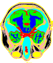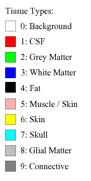

Anatomical model. This model was used by the MRI Simulator to generate the MRI data sets with different noise levels.
|
|
|
|---|---|
|
Anatomical model. This model was used by the MRI Simulator to generate the MRI data sets with different noise levels. |
|
|
|
|
|
|
|---|---|---|---|
|
|
|
|
|
|
|
|
|
|
|
|
|
|
|
|
MRI data sets with 3%, 5%, 7% and 9% noise. |
Segmented image |
Variance image (of the segmented image) |
Variance image of the anatomical model |
|
|
|
|
|
|---|---|---|---|
|
|
|
|
|
|
|
|
|
|
|
|
|
|
|
|
MRI data sets with 3%, 5%, 7% and 9% noise. |
Segmented image |
Variance image (of the segmented image) |
Variance image of the anatomical model |
Overall Error Diagram for 3% Noise MRI |
|
|
|---|
|
x-axis: number of segmented features; y-axis: error rate (and variance from 50 segmentation results). |
Overall Error Diagram for 5% Noise MRI |
|
|
|---|
|
x-axis: number of segmented features; y-axis: error rate (and variance from 50 segmentation results). |
Overall Error Diagram for 7% Noise MRI |
|
|
|---|
|
x-axis: number of segmented features; y-axis: error rate (and variance from 50 segmentation results). |
Overall Error Diagram for 9% Noise MRI |
|
|
|---|
|
x-axis: number of segmented features; y-axis: error rate (and variance from 50 segmentation results). |
Mutual Information Diagram for 3% Noise MRI |
|
|
|---|
|
x-axis: Gibbs potential ß; y-axis: Mutual Information (and variance from 50 segmentation results) |
Mutual Information Diagram for 5% Noise MRI |
|
|
|---|
|
x-axis: Gibbs potential ß; y-axis: Mutual Information (and variance from 50 segmentation results) |
Mutual Information Diagram for 7% Noise MRI |
|
|
|---|
|
x-axis: Gibbs potential ß; y-axis: Mutual Information (and variance from 50 segmentation results) |
Mutual Information Diagram for 9% Noise MRI |
|
|
|---|
|
x-axis: Gibbs potential ß; y-axis: Mutual Information (and variance from 50 segmentation results) |
![[Image of CS Logo]](https://www.cosy.sbg.ac.at/~held/outline_cga_lab_s.gif) |
Copyright © 2025
Martin Held, held@cs.plus.ac.at.
All rights reserved.
Impressum and Disclaimer. file last modified: Thursday, 27-Feb-2020 07:29:07 CET |
![[Image of CS Logo]](https://www.cosy.sbg.ac.at/~held/outline_cga_lab_s.gif) |